
How to Differentiate Skin Spots? A Scientific and Clinical Approach
What is a Skin Lesion and Spot?
Skin lesions are abnormal skin changes that appear different from the surrounding tissue. These changes can manifest as color changes, swelling, or scars. While most lesions are benign, some may indicate serious diseases. The distinction between lesions and spots is critically important in practice. A lesion refers to any pathological change in tissue integrity, while a spot typically indicates changes in pigmentation.
Why are Skin Spots Important?
Clinically accurate differential diagnosis is vital. Early diagnosis is crucial, especially for lesions with potential for malignant transformation. Skin spots should be evaluated from both cosmetic and medical perspectives. Sun exposure, hormonal changes, and genetic factors play a major role in the formation of spots.
How are Skin Lesions Classified?
What are Primary Lesions?
Primary lesions are the initial pathological changes that occur in the skin. These lesions are direct products of the disease process and have not undergone secondary changes.
Lesion Type | Definition | Clinical Feature |
Macule | Flat color change | Not felt on the skin surface, shows only color difference |
Papule | Small bump | Firm structure, acne-like swelling |
Nodule | Mass located in the skin | Palpable mass exceeding one centimeter |
Vesicle | Fluid-filled bubble | Small in diameter, filled with clear fluid |
Pustule | Lesion filled with pus | Contains white or yellow pus |
What Are Secondary Lesions?
Secondary lesions arise as a result of the evolution of primary lesions. Itching, injury, or an underlying disease process triggers these lesions.
Lesion Type | Definition | Cause of Formation |
Crust (Scab) | Dried fluid | Occurs after a wound or vesicle |
Scar | Fibrous tissue | Permanent tissue formed after wound healing |
Ulcer | Open wound | Occurs due to loss of epidermis and dermis |
Atrophy | Thin skin | Thinning due to tissue loss |
Erythema | Redness | Occurs due to the dilation of blood vessels |
How to Distinguish Skin Spots by Color Characteristics?
What is Hyperpigmentation?
Hyperpigmentation refers to dark spots that occur due to an increase in melanin in certain areas of the skin. Melanin biosynthesis is a complex enzymatic process that occurs in melanocytes. The enzyme tyrosinase plays a critical role in this process. Tyrosine is converted first to L-DOPA and then to dopachrome under the influence of tyrosinase. As a result of these reactions, melanin polymer is formed (Özçelik, 2005).
What is Hypopigmentation?
Hypopigmentation is the formation of lighter areas on the skin due to a decrease in melanin production or loss of melanocytes. Vitiligo, post-inflammatory hypopigmentation, and albinism are among the main causes.
What is Erythema?
Erythema is a lesion that appears as redness on the skin. It occurs due to the dilation of blood vessels. Infections, allergies, and inflammatory processes lead to erythema.
What are the Common Types of Skin Spots?
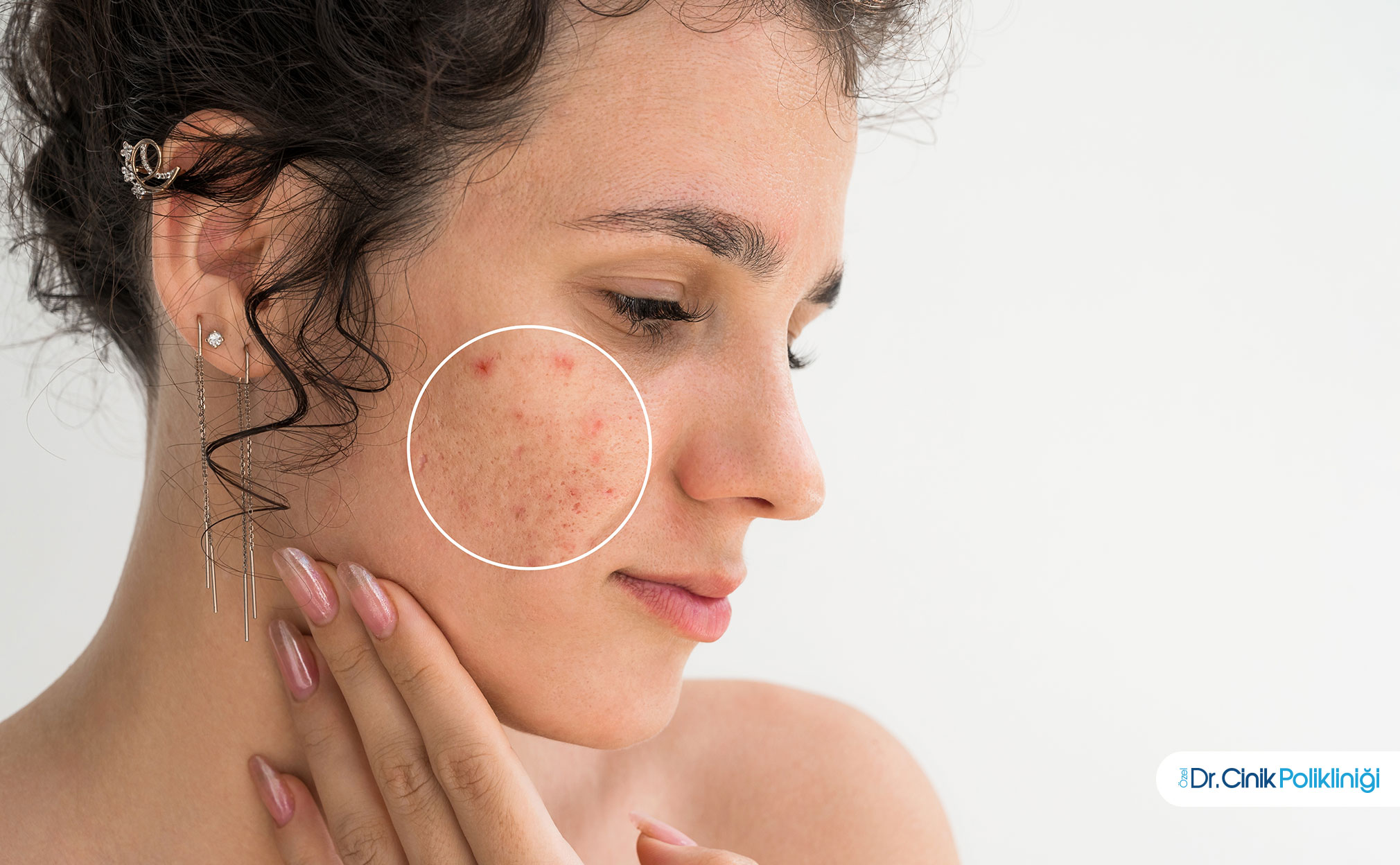
How to Recognize Acne and Its Subtypes?
Acne vulgaris is a multifactorial, inflammatory disease of the pilosebaceous unit. Four classic mechanisms play a role in its pathogenesis: increased sebum production, bacterial colonization, follicular hyperkeratinization, and inflammation (Kesikoğlu and Çınar, 2023).
Comparison of Acne Lesions:
Lesion Type | Appearance | Pathophysiology |
Comedo (Blackhead) | Open pore, black-tipped | Follicle blockage, oxidized oil |
Comedo (Whitehead) | Closed pore, white-tipped | Follicle blockage, anaerobic environment |
Papule | Red, raised, non-inflammatory | Beginning of inflammatory response |
Pustule | Filled with pus, white-tipped | Bacterial colonization, neutrophil infiltration |
Nodule | Deep, painful mass | Severe inflammation, tissue damage |
Cystic Acne | Large mass filled with fluid | Chronic inflammation, high risk of scarring |
How to Differentiate Pigmentation Spots?
What are Sun Spots (Lentigo)?
Solar lentigo are flat, dark brown spots that occur as a result of chronic sun exposure. They are commonly seen in middle-aged and older groups in sun-exposed areas (face, back of hands, forearms).
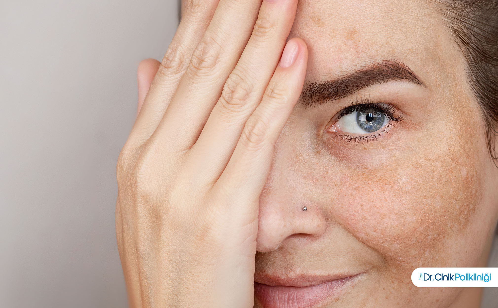
What is Melasma?
Melasma is a common hyperpigmentation disorder resulting from excessive production of melanin pigment in the skin. It is usually seen on the face, particularly on the forehead, cheeks, bridge of the nose, and upper lip. It is much more common in women than in men, with a reported ratio of 39 female patients for every male patient (Mercan-Bozkurt, 2024).
Comparison of Melasma and Sun Spots:
Feature | Melasma | Solar Lentigo |
Distribution | Symmetric, centered on the face | Asymmetric, sun-exposed areas |
Color | Brown-gray | Smooth brown |
Border | Irregular | Smooth |
Hormonal Relationship | Strong (pregnancy, birth control) | None |
Skin Type | III-V more common | All types |
What is Post-Inflammatory Hyperpigmentation?
Post-inflammatory hyperpigmentation refers to dark marks that appear on the skin following acne, wounds, burns, or irritation. The increase in melanocyte activity after inflammation leads to increased melanin production. It is particularly more pronounced in darker skin types.
How are Benign Lesions Recognized?
What is a Nevus?
A nevus is a benign lesion that results from the abnormal proliferation of melanocytes. It is classified into three main types: intradermal, junctional, and compound nevus (Özgür, 2024).
Types of Nevus:
Type | Location | Clinical Appearance | Malignant Potential |
Intraderma Nevus | Within the dermis | Raised, brown, may be hairy | None |
Junctional Nevus | Basal membrane | Flat, hairless, shades of brown | Very low |
Compound Nevus | Both epidermis and dermis | Raised, may contain hair | Low |
Dysplastic Nevus | Irregularly defined | Large, variegated colors | High (10 times increased risk) |
What are Freckles and Skin Tags?
Freckles are pigmented lesions that occur due to an abnormal number of melanin granules produced by normal amounts of melanocytes. There is no possibility of malignant transformation. Skin tags are soft, hanging benign fibroepithelial polyps.
How to Recognize Potential Malignant Lesions?
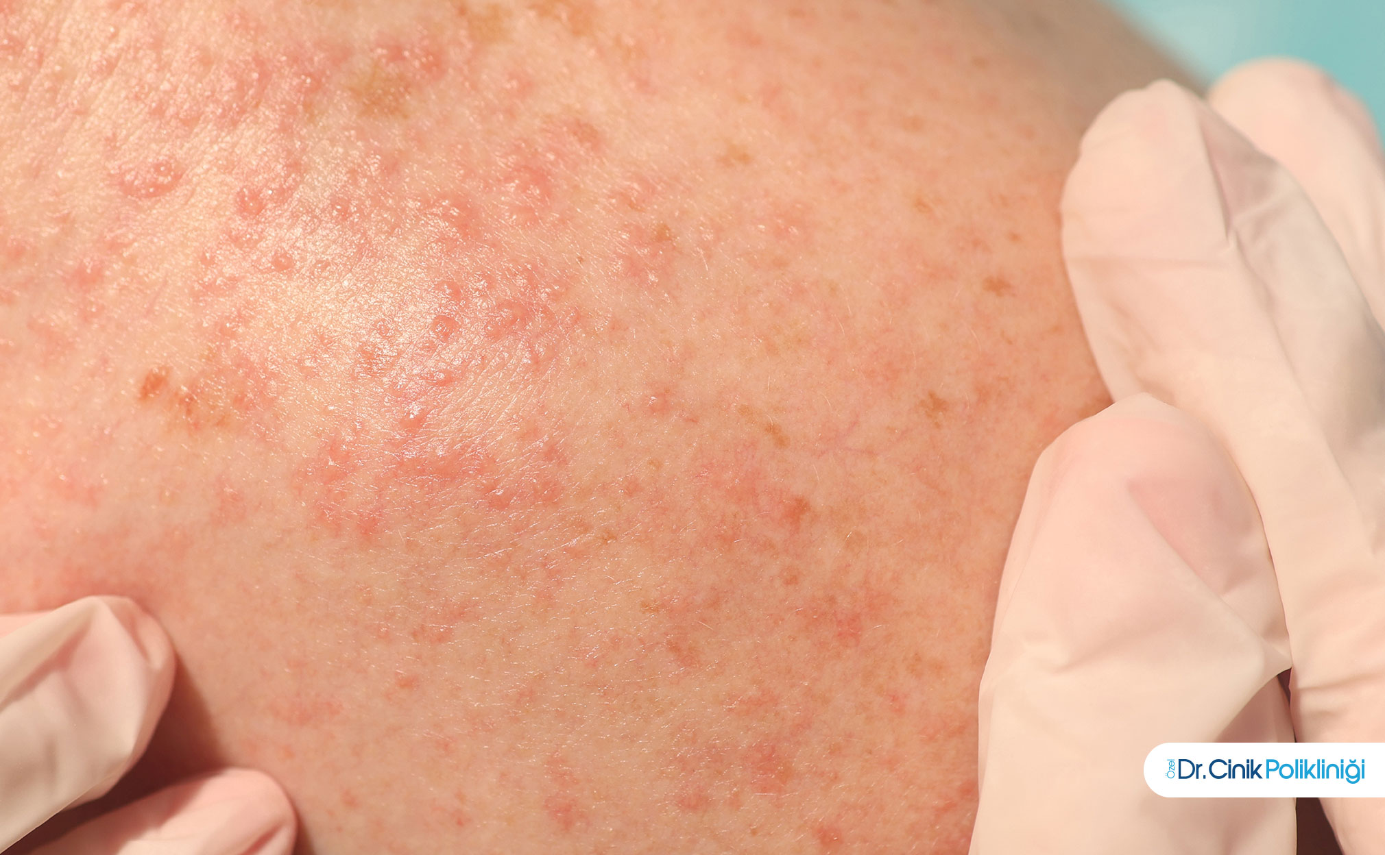
What is Actinic Keratosis?
Actinic keratosis is a precancerous skin lesion that develops as a result of prolonged exposure to sunlight. It arises due to long-term damage caused by ultraviolet rays on the skin. It has a 20% potential to transform into squamous cell carcinoma (Skin Cancer Association, 2024).
Characteristics of Actinic Keratosis:
Occurs in sun-exposed areas (face, scalp, forearm, back of the hand)
Rough, dry, scaly surface
Redness and sometimes slight elevation
More common in Fitzpatrick skin types I-II
How to Identify Lesions Suspicious for Melanoma?
Melanoma is an aggressive skin cancer that results from the malignant proliferation of melanocytes. Early diagnosis determines the success of treatment. The ABCDE criteria are the standard assessment tool for recognizing lesions suspicious for melanoma (Hira, 2026).
ABCDE Criteria:
Criterion | Description | Risk Sign |
A - Asymmetry | One benign half does not resemble the other | Asymmetrical structure |
B - Border | Smoothness of edges | Irregular, scalloped, indistinct border |
C - Color | Color distribution | Multicolored (brown, black, red, blue) |
D - Diameter | Size | Greater than 6 mm |
E - Evolution | Change | Change in size, color, or shape |
What Are the Differences Between Acne, Spots, and Scars?
How to Differentiate Between Active Acne and Acne Spots?
Active acne refers to lesions where the inflammatory process is ongoing. Acne spots are pigment changes that remain after inflammation. Active acne can be painful, red, and swollen; spots are flat and present as color changes.
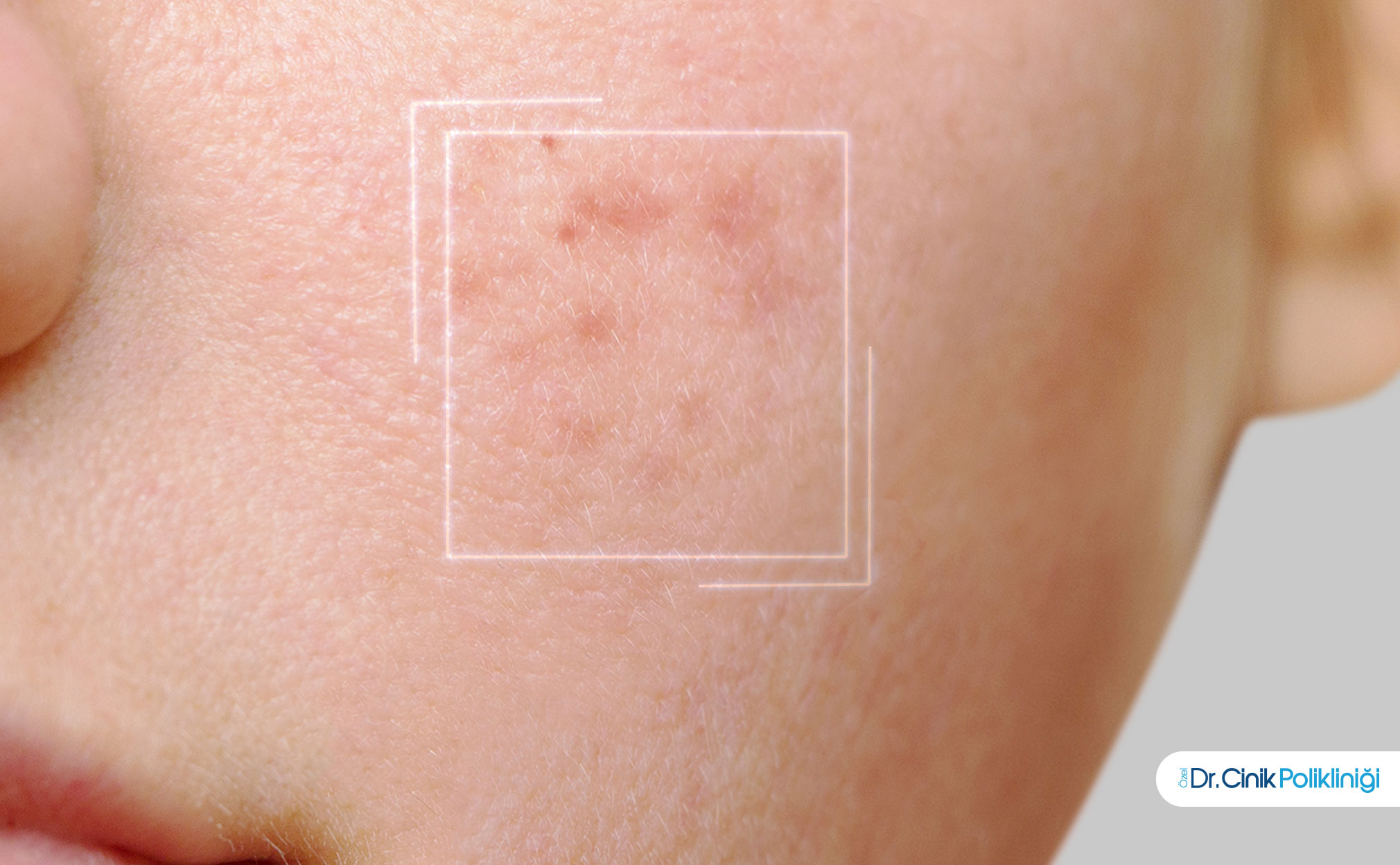
What Is the Difference Between a Spot and a Scar?
A spot is a pigment-based change. There is no tissue damage. A scar is a permanent change resulting from tissue damage and fibrosis. Scars indicate deep tissue loss or excess.
Comparison Table:
Feature | Spot (Pigment) | Scar |
Structure | Flat, no tissue change | Raised or depressed, tissue change present |
Color | Brown, gray | Pink, white, red |
Touch | Normal skin texture | Hard or soft abnormal tissue |
Healing | With pigment regulating treatment | Limited healing, revision needed |
What Are the Causes of Skin Spot Formation?
What Are Endogenous Factors?
Genetic predisposition plays a fundamental role in the formation of spots. The risk increases by 30% in individuals with a family history of melasma. Hormonal changes, especially during pregnancy and the use of birth control pills, increase melanin synthesis. Estrogen induces melanogenesis by increasing tyrosinase expression through nuclear receptors (Erkekoğlu et al., 2022).
What Are Exogenous Factors?
Ultraviolet exposure is the strongest trigger for melanin synthesis. UV rays induce melanogenesis through melanocyte-stimulating hormone (MSH) released from keratinocytes and endothelin-1. Cosmetic products and environmental factors also contribute to the formation of spots.
What Are Microbial and Immunological Factors?
Cutibacterium acnes (formerly known as Propionibacterium acnes) plays a central role in acne pathogenesis. This bacterium proliferates in an anaerobic environment within the follicle and leads to the release of inflammatory cytokines. P. acnes causes cytokine production by carrying TLR ligands and enhances inflammation (Perçin, 2023).
What Are the Diagnostic Methods for Skin Spots?
How Is Clinical Evaluation Conducted?
Visual inspection is the first step in diagnosis. The color, size, shape, border characteristics, and palpation findings of the lesion are recorded. A systematic examination requires creating a body map.
What Is Dermatoscopy?
Dermatoscopy is a non-invasive method that allows the examination of lesions on the skin surface using a dermatoscope, which is a special magnification and illumination system. Lesions are examined in detail with 10-70 times magnification. This method facilitates capturing truly risky lesions while reducing unnecessary biopsies (Filiz Özgür, 2024).
When Is a Biopsy Necessary?
Histopathological examination confirms the diagnosis in suspicious lesions. There is an indication for biopsy in lesions that meet the ABCDE criteria and show melanoma-like features in dermatoscopy.
When Should You See a Doctor?
evaluation is urgently recommended in the following situations:
Rapidly growing lesions
Moles showing color changes
Bleeding or non-healing wounds
Itchy, painful lesions
Changes that meet the ABCDE criteria
How Are Skin Spots Treated?
What Are the Medical Treatment Options?
Topical agents are the first step in the treatment of hyperpigmentation. Hydroquinone inhibits melanin synthesis by inhibiting the tyrosinase enzyme. Retinoids regulate pigment distribution by accelerating keratinocyte turnover.
What are Procedures?
Procedure | Indication | Mechanism |
Deep pigment spots | Selective photothermolysis | |
Superficial pigmentation | Controlled epidermal renewal | |
Cryotherapy | Actinic keratosis | Cell destruction with liquid nitrogen |
Photodynamic Therapy | Premalignant lesions | Photosensitizer and light |
What are Preventive Approaches?
The use of sunscreen prevents the formation of spots and the worsening of existing spots. Sunscreens containing iron oxide that block not only UVA and UVB but also visible light significantly reduce the recurrence of melasma (Erkekoğlu et al., 2022).
Frequently Asked Questions
How are skin spots distinguished?
Skin spots are classified according to color, texture, distribution, and evolution characteristics. Hyperpigmentation refers to dark spots, while hypopigmentation refers to light-colored spots. Flat lesions are called macules, while raised lesions are referred to as papules and nodules.
Which skin lesions are dangerous?
Rapidly growing lesions that show color change, bleed, or meet the ABCDE criteria are dangerous. Actinic keratosis and dysplastic nevus carry malignant potential.
How to distinguish acne scars from sun spots?
An acne scar forms at the site of a previous acne lesion and is usually dark brown. A sun spot (solar lentigo) appears as well-defined, flat spots in sun-exposed areas.
Do skin spots go away on their own?
Some spots (especially post-inflammatory hyperpigmentation) may fade over time. However, melasma and sun spots can be permanent and require treatment.
References
Erkekoğlu, P., and colleagues. "Current Approaches and Possible Toxic Effects in the Treatment of Melasma." Journal of Hacettepe University Faculty of Pharmacy, vol. 42, no. 2, 2022, pp. 105-120.
Hira, Hüseyin. "What is a Mole Examination? Dermatoscopy for Melanoma Screening and Early Diagnosis." 2026.
Kesikoğlu, Ayten Ferahbaş, and Salih Levent Çınar. "Overview of the Etiopathogenesis of Acne Vulgaris." Turkderm, 2023.
Mercan-Bozkurt, Nurhal. "Sun Spots and Melasma." NMB Clinic, 2024.
Özçelik, B. "Tyrosinase Inhibitors." Istanbul University Institute of Science, 2005.
Özgür, Filiz. "Dermatoscopy." Dr. Filiz Özgür Çavuş , 2024.
Perçin, Duygu. "The Acne Book." Kütahya Health Sciences University Faculty of Medicine, 2023.
Turkey Skin Cancer Association. "Lesions that are Precursors (Premalignant) to Skin Cancer." 2024.
Get in touch with our expert team
Related Posts


